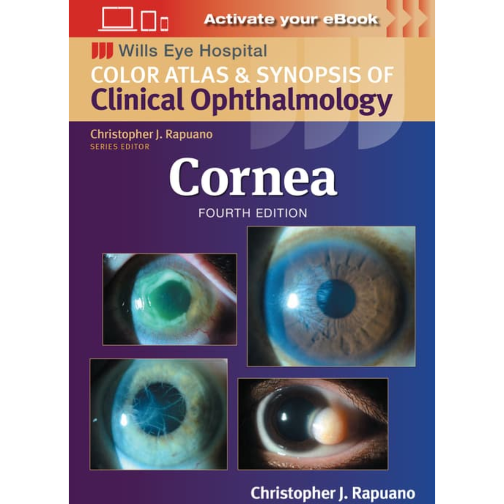
Cornea (Wills Eye Institute Atlas Series)
9781975214975
-
₪404.00₪449.00
אזל המלאי
פריט זה ניתן על ידי קרדיטים.
לחצו על "הוספה לעגלה" להמשך
לחצו על "הוספה לעגלה" להמשך
אזל המלאי עבור /
-
זמן אספקה ותנאי רכישההערות:
• זמן אספקה: הזמנות בהן כל הספרים זמינים במלאי - זמן אספקה – כ- 5 ימי עסקים (למעט אזורים חריגים בהם ייתכן עיכוב נוסף).
ספרים שאינם זמינים במלאי: זמן אספקה כ- 14 -30 ימי עסקים בהתאם למלאי במחסני המו"ל בחו"ל - הודעה תימסר ללקוח.
• הזמנה במשקל כולל של עד 14 קילו ישלחו ללקוח באמצעות חברת שליחויות עם שליח עד הבית (בישובים מסוימים המסירה תתבצע למרכז חלוקת הדואר המקומי)
• במידה וקיים עיכוב במשלוח ההזמנה או חוסר במלאי הספרים תשלח הודעה ללקוח.
• במידה ויבחר הלקוח עקב עיכוב במשלוח כנ"ל לבטל הזמנתו ויודיע על כך לידע, ידע מתחייבת לזכות החיוב.
• במידה ויתברר כי הספרים אזלו מהמלאי ולא ניתן לספקם - תשלח הודעה ללקוח.
• האיסוף העצמי ממשרדי ידע יבוצע רק לאחר הודעה ללקוח שההזמנה מוכנה לאיסוף.
דמי משלוח:
ניתן לבחור: 1. איסוף עצמי - ללא תשלום
2. משלוח עד הבית
Cornea \ Christopher J Rapuano MD
4rd edition, 2024
Developed at Philadelphia’s world-renowned Wills Eye Hospital, the Color Atlas and Synopsis of Clinical Ophthalmology series covers the most clinically relevant aspects of ophthalmology in a highly visual, easy-to-use format. Vibrant, full-color photos and a consistent outline structure present a succinct, high-yield approach to the seven topics covered by this popular series: Cornea, Retina, Glaucoma, Oculoplastics, Neuro-Ophthalmology, Pediatrics, and Uveitis. This in-depth, focused approach makes each volume an excellent companion to the larger Wills Eye Manual as well as a practical stand-alone reference for students, residents, and practitioners in every area of ophthalmology.
The updated Cornea volume includes:
- Expert guidelines for the differential diagnosis and treatment of cornea diseases seen by the ophthalmic resident, general ophthalmologist, and cornea specialist
- Up-to-date information on infections and complications of corneal surgeries
- More than 450 high-quality photographs of important corneal, anterior segment, and external diseases, many new and updated for this fourth edition
- Revised coverage of the clinical features of key cornea and external eye diseases, diagnostic tests, differential diagnoses, and treatment
- 434 additional full-color photographs and drawings further illustrate pathology and therapeutics described in the text
מוצרים קשורים
-
יח'יח'
תודה על השיתוף
קיבלתם הנחת שיתוף מיוחדת! על מנת להינות מהנחה זו עליכם להוסיף את הפריט לעגלת הקניות בכפתור הוספה לעגלה.
הצטרפו לרשימת המתנה לחזרה למלאי
הצטרפות לרשימת ההמתנה בוצעה בהצלחה.
אנו נשלח אליכם מייל כאשר הפריט יחזור למלאי.


