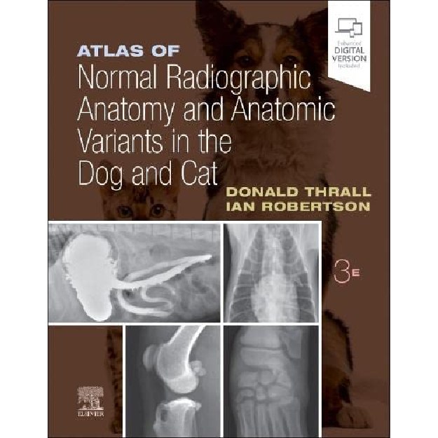
Atlas of Normal Radiographic Anatomy and Anatomic Variants in the Dog and Cat
9780323796156
-
₪926.00₪1,030.00
אזל המלאי
פריט זה ניתן על ידי קרדיטים.
לחצו על "הוספה לעגלה" להמשך
לחצו על "הוספה לעגלה" להמשך
אזל המלאי עבור /
-
זמן אספקה ותנאי רכישההערות:
• זמן אספקה: הזמנות בהן כל הספרים זמינים במלאי - זמן אספקה – כ- 5 ימי עסקים (למעט אזורים חריגים בהם ייתכן עיכוב נוסף).
ספרים שאינם זמינים במלאי: זמן אספקה כ- 14 -30 ימי עסקים בהתאם למלאי במחסני המו"ל בחו"ל - הודעה תימסר ללקוח.
• הזמנה במשקל כולל של עד 14 קילו ישלחו ללקוח באמצעות חברת שליחויות עם שליח עד הבית (בישובים מסוימים המסירה תתבצע למרכז חלוקת הדואר המקומי)
• במידה וקיים עיכוב במשלוח ההזמנה או חוסר במלאי הספרים תשלח הודעה ללקוח.
• במידה ויבחר הלקוח עקב עיכוב במשלוח כנ"ל לבטל הזמנתו ויודיע על כך לידע, ידע מתחייבת לזכות החיוב.
• במידה ויתברר כי הספרים אזלו מהמלאי ולא ניתן לספקם - תשלח הודעה ללקוח.
• האיסוף העצמי ממשרדי ידע יבוצע רק לאחר הודעה ללקוח שההזמנה מוכנה לאיסוף.
דמי משלוח:
ניתן לבחור: 1. איסוף עצמי - ללא תשלום
2. משלוח עד הבית
Atlas of Normal Radiographic Anatomy and Anatomic Variants in the Dog and Cat \ By (author) Donald E. Thrall , By (author) Ian D. Robertson
3rd edition, 2022
Make accurate diagnoses and achieve successful treatment outcomes with this highly visual, comprehensive atlas! Featuring a substantial number of new high-contrast images, Atlas of Normal Radiographic Anatomy and Anatomic Variants in the Dog and Cat, 3rd Edition, provides an in-depth look at both normal and non-standard subjects, along with demonstrations of proper technique and image interpretations. Expert authors Donald E. Thrall and Ian D. Robertson describe a wider range of "normal" as compared to other books - not only showing standard dogs and cats, but also non-standard subjects such as overweight and underweight pets and animals with breed-specific variations. Each body part is put into context with a description that helps explain why a structure appears as it does in radiographs, enabling you to appreciate variations of normal based on an understanding of basic radiographic principles.
UPDATED! Brief descriptive text and explanatory legends accompany all images to help put concepts into the proper context.
High-quality digital images provide excellent contrast resolution and better visibility of normal structures to facilitate making accurate diagnoses.
In-depth coverage of patient positioning and radiographic exposure guidelines assists in producing the very best results.
NEW! Expanded coverage of the neonatal and juvenile subject includes additional radiographic examples.
NEW! Additional material on the normal appearance of some of the more common special procedures performed in private practice includes barium esophagram, barium gastrointestinal study, and positive contrast cystogram.
NEW! Coverage of shoulder arthrography illustrates the normal expected location of the joint capsule.
NEW and UPDATED! Radiographic images of normal or standard prototypical animals are supplemented by images of non-standard subjects exhibiting breed-specific differences, physiologic variants, or common congenital malformations.
NEW! Enhanced ebook, included with the purchase of a new print copy of the book, provides online access to a fully searchable version of the text and makes its content available on various devices.
מוצרים קשורים
-
יח'יח'יח'
תודה על השיתוף
קיבלתם הנחת שיתוף מיוחדת! על מנת להינות מהנחה זו עליכם להוסיף את הפריט לעגלת הקניות בכפתור הוספה לעגלה.
הצטרפו לרשימת המתנה לחזרה למלאי
הצטרפות לרשימת ההמתנה בוצעה בהצלחה.
אנו נשלח אליכם מייל כאשר הפריט יחזור למלאי.


