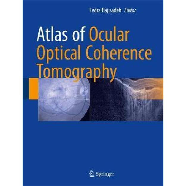
Atlas of Ocular Optical Coherence Tomography
9783319667560
-
₪1,188.00
אזל המלאי
פריט זה ניתן על ידי קרדיטים.
לחצו על "הוספה לעגלה" להמשך
לחצו על "הוספה לעגלה" להמשך
אזל המלאי עבור /
-
זמן אספקה ותנאי רכישההערות:
• זמן אספקה: הזמנות בהן כל הספרים זמינים במלאי - זמן אספקה – כ- 5 ימי עסקים (למעט אזורים חריגים בהם ייתכן עיכוב נוסף).
ספרים שאינם זמינים במלאי: זמן אספקה כ- 14 -45 ימי עסקים בהתאם למלאי במחסני המו"ל בחו"ל - הודעה תימסר ללקוח.
• הזמנה במשקל כולל של עד 14 קילו ישלחו ללקוח באמצעות חברת שליחויות עם שליח עד הבית (בישובים מסוימים המסירה תתבצע למרכז חלוקת הדואר המקומי)
• במידה וקיים עיכוב במשלוח ההזמנה או חוסר במלאי הספרים תשלח הודעה ללקוח.
• במידה ויבחר הלקוח עקב עיכוב במשלוח כנ"ל לבטל הזמנתו ויודיע על כך לידע, ידע מתחייבת לזכות החיוב.
• במידה ויתברר כי הספרים אזלו מהמלאי ולא ניתן לספקם - תשלח הודעה ללקוח.
• האיסוף העצמי ממשרדי ידע יבוצע רק לאחר הודעה ללקוח שההזמנה מוכנה לאיסוף.
דמי משלוח:
ניתן לבחור: 1. איסוף עצמי - ללא תשלום
2. משלוח עד הבית
Atlas of Ocular Optical Coherence Tomography \ Fedra Hajizadeh
2018
This book provides a collection of optical coherence tomographic (OCT) images of various diseases of posterior and anterior segments. It covers the details and issues of diagnostic tests based on OCT findings which are crucial for ophthalmologists to understand in their clinical practice. Throughout the chapters all aspects of this non-invasive, popular imaging technique, known for ingenuity and accuracy, is clearly illustrated.
Atlas of Ocular Optical Coherence Tomography has been categorized into eleven sections, discussing and illustrating distinct OCT features, as well as showing other image modalities such as fluorescein angiography, fundus autofluorescence, perimetry and laboratory examination. This book also covers choroidal pathologies and vitreous abnormalities. The last section has been allocated to anterior segment disease, including cornea, angle, iris and conjunctival abnormalities. Above all, the numerous images, and detailed descriptions of diseases, make this book an essential guide for general ophthalmologists and ophthalmology residences.
מוצרים קשורים
-
יח'יח'יח'
תודה על השיתוף
קיבלתם הנחת שיתוף מיוחדת! על מנת להינות מהנחה זו עליכם להוסיף את הפריט לעגלת הקניות בכפתור הוספה לעגלה.
הצטרפו לרשימת המתנה לחזרה למלאי
הצטרפות לרשימת ההמתנה בוצעה בהצלחה.
אנו נשלח אליכם מייל כאשר הפריט יחזור למלאי.
