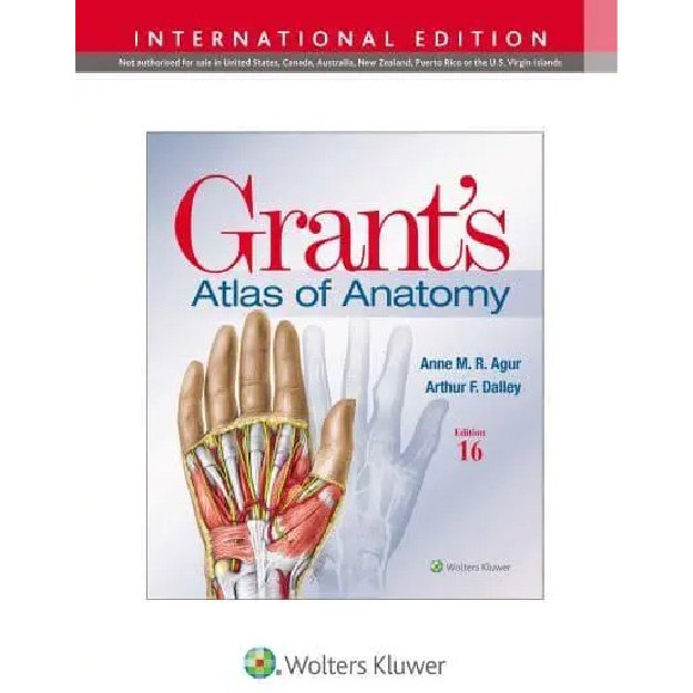
Grant's Atlas of Anatomy 16th Edition
9781975193447
-
₪485.00₪540.00
אזל המלאי
פריט זה ניתן על ידי קרדיטים.
לחצו על "הוספה לעגלה" להמשך
לחצו על "הוספה לעגלה" להמשך
אזל המלאי עבור /
-
זמן אספקה ותנאי רכישההערות:
• זמן אספקה: הזמנות בהן כל הספרים זמינים במלאי - זמן אספקה – כ- 5 ימי עסקים (למעט אזורים חריגים בהם ייתכן עיכוב נוסף).
ספרים שאינם זמינים במלאי: זמן אספקה כ- 14 -45 ימי עסקים בהתאם למלאי במחסני המו"ל בחו"ל - הודעה תימסר ללקוח.
• הזמנה במשקל כולל של עד 14 קילו ישלחו ללקוח באמצעות חברת שליחויות עם שליח עד הבית (בישובים מסוימים המסירה תתבצע למרכז חלוקת הדואר המקומי)
• במידה וקיים עיכוב במשלוח ההזמנה או חוסר במלאי הספרים תשלח הודעה ללקוח.
• במידה ויבחר הלקוח עקב עיכוב במשלוח כנ"ל לבטל הזמנתו ויודיע על כך לידע, ידע מתחייבת לזכות החיוב.
• במידה ויתברר כי הספרים אזלו מהמלאי ולא ניתן לספקם - תשלח הודעה ללקוח.
• האיסוף העצמי ממשרדי ידע יבוצע רק לאחר הודעה ללקוח שההזמנה מוכנה לאיסוף.
דמי משלוח:
ניתן לבחור: 1. איסוף עצמי - ללא תשלום
2. משלוח עד הבית
Grant's Atlas of Anatomy \ A. M. R. Agur, Arthur F. Dalley, J. C. Boileau Grant
16th Edition, 2024
Illustrations drawn from real specimens, presented in surface-to-deep dissection sequence, set Grant's Atlas of Anatomy apart as the most accurate illustrated reference available for learning human anatomy and referencing in dissection lab. A recent edition featured re-colorization of the original Grant's Atlas images from high-resolution scans, also adding a new level of organ luminosity and tissue transparency. The dissection illustrations are supported by descriptive text legends with clinical insights, summary tables, orientation and schematic drawings, and medical imaging.
Illustrations drawn from real specimens, presented in surface-to-deep dissection sequence, set Grant's Atlas of Anatomy apart as the most accurate illustrated reference available for learning human anatomy and referencing in dissection lab. A recent edition featured re-colorization of the original Grant's Atlas images from high-resolution scans, also adding a new level of organ luminosity and tissue transparency. The dissection illustrations are supported by descriptive text legends with clinical insights, summary tables, orientation and schematic drawings, and medical imaging.
- Renowned, high-resolution, dynamically colored illustrations organized in dissection sequence enable the formation of 3D constructs for each body region and provide detailed, realistic reference during dissection.
- Tables detail muscles, vessels, and other anatomic information in an easy-to-use format ideal for review and study.
- Enhanced medical imaging includes more than 100 clinically significant MRIs, CT images, ultrasound scans, and corresponding orientation drawings to help students confidently apply the laboratory experience to clinical rotations.
- Color schematic illustrations reinforce the relationships of structures and anatomical concepts in vibrant detail.
מוצרים קשורים
-
יח'יח'יח'
תודה על השיתוף
קיבלתם הנחת שיתוף מיוחדת! על מנת להינות מהנחה זו עליכם להוסיף את הפריט לעגלת הקניות בכפתור הוספה לעגלה.
הצטרפו לרשימת המתנה לחזרה למלאי
הצטרפות לרשימת ההמתנה בוצעה בהצלחה.
אנו נשלח אליכם מייל כאשר הפריט יחזור למלאי.


